ST. JOHN'S, N.L. — Everyone wants a PET scanner, but not everyone knows what they do.
In a nutshell, they pinpoint problem spots in a patient's body so a specialist can consider treatment options. While they are used for heart or neurological anomalies, they are most commonly used in seeking out cancerous tumours.
Unlike a CT (computerized tomography) scanner, which uses X-rays to produce a 3-D image of the body, a PET (positron emission tomography) scanner detects tiny “particles” of light, or protons, that are emitted by a radioactive substance injected into the body.
The substance used for PET scans emits positrons, which are no bigger than electrons. The amount of radiation is very low and safe. In fact, when they combine with electrons in the body, they are destroyed and give off the tiny specks of light that the machine picks up.
The positrons injected are often attached to molecules of sugar called fluorodeoxyglucose (FDG). Why sugar? Because cancer cells are more aggressive and grow at a faster rate, consuming sugar in the process. The radioactive sugar tends to accumulate around these cells.
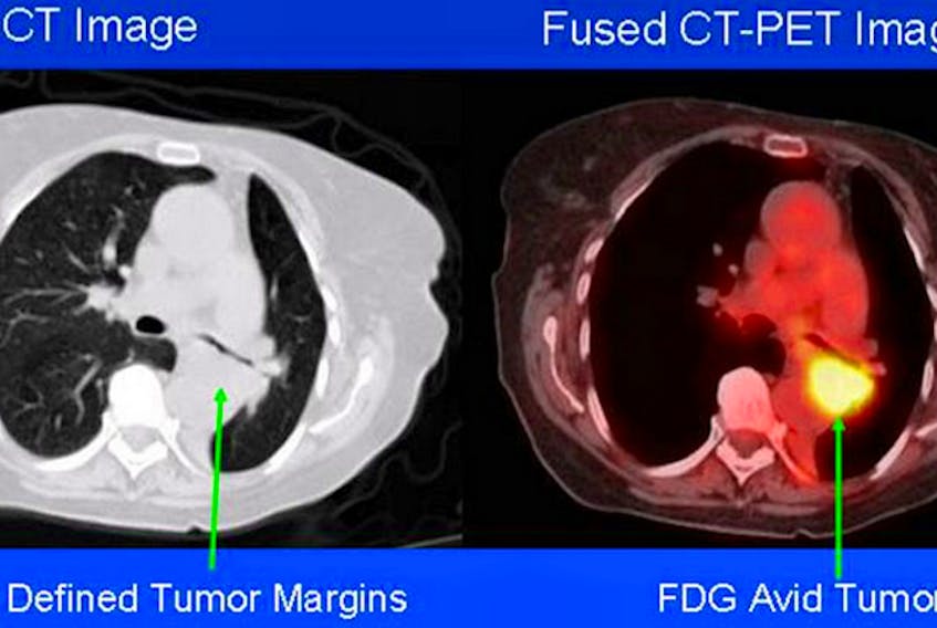
The result is an eerie glow that shows up on the screen, and the intensity of that glow can even indicate how aggressive the cancer is.
Often, a PET scan is done in conjunction with a CT scan and the two merged through computer software. This gives a better framework to pinpoint the glowing tumour.
Similar to a PET scanner is a SPECT scanner (single-photon emission computerized tomography), except the isotopes used in this case emit gamma rays. Their purpose is to show how a patient's organs are working. They can show how blood flows to the heart and which areas of the brain are more active or less active.